New academic year, round 7 of Kavli INsD ‘3-min researcher updates’ and still going strong.
New Academic Year, Round 7 of Kavli INsD ‘3-min Researcher Updates’ and Still Going Strong
To foster interdisciplinarity and understanding of each other's fields and methodologies, our researchers at Kavli INsD continued to meet up informally during October 2023 to share brief 3-minute updates on their work.
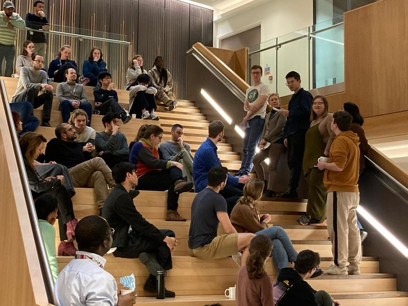
Through this exchange, we aim to address significant health challenges such as antimicrobial resistance, brain and mental health, infectious diseases, and malaria. Additionally, we strive to develop innovative instrumentation that harnesses the analytical capabilities of the physical sciences to investigate cellular processes. Together, we are committed to making substantial contributions in these areas.
We are pleased to share our researcher’s presentation summaries below.
|
Physiology, Anatomy and genetics |
Clinical Neuroscience |
Biochemistry |
Physics |
Chemistry |
|
Dr Stewart Humble |
Dr Suman Dutta |
Dr Sarah Silk |
Dr Martin Rieu |
Dr Linlin Zhang |
|
Benjamin Breant (DPhil Candidate) |
Tatiana Wilson (DPhil candidate) |
Dr Emma Silvester |
Dr Jagadish Prasad Hazra |
Dr Abraham Oluwole |
|
Dr David López Martínez |
Dr Vojta Pražák |
Dr Stelios Chatzimichail |
Dr Tim Esser |

Dr Jagadish Prasad Hazra
Towards Sequence dependent kinetics using a novel kinetics based single molecule sequencing.
Transcription begins with the sequence-dependent binding of the RNA polymerase holoenzyme to the promoter DNA, followed by short pauses and the synthesis of short RNA transcripts, termed abortive initiation, before successful initiation. These steps are controlled by the specific sequences of the promoter. Despite extensive research, understanding how sequence affects transcription kinetics has remained elusive. To address this gap, a platform connecting DNA genotype to kinetic phenotype at the single-molecule level is being developed. In the first step, we created a high-throughput DNA-hybridization-based single-molecule DNA sequencing method. Machine learning-based analytical approaches provide higher accuracy and faster analysis. The second step involves studying the propensity of different DNA sequences at specific positions in the promoter to cause initiation pausing. This research offers insights into gene expression regulation and transcription initiation mechanisms.

Dr Linlin Zhang
Guiding Antiviral Discovery against Filoviruses via Glycosylation Analysis of Viral Spikes
I am fascinated by the mechanisms of viral-host interaction, as the glycosylation of spike fusion glycoproteins is an important functional feature of enveloped viruses and are common targets for therapeutic intervention and vaccine design. My current research is seeking to understand how glycans influence the structural dynamics and interactions, specifically viruses that pose the greatest threat to global health – Ebola (EBOV) and Marburg (MARV). Part of my research entails mapping the site-specific glycan structures on EBOV and MARV GP glycoproteins produced in various cell lines and correlating changes in glycosylation with binding affinities to glycan-dependent EBOV/MARV receptors. Significantly, the protocols, methods and strategies developed from this research can potentially be applied for a rapid response to the re(emerging) enveloped viruses in the future.

Dr David López Martínez
iPSC cells, dopaminergic neurons, and ubiquitin system to elucidate Parkinson's disease.
I am studying the molecular mechanism of a-synuclein degradation because the a-synuclein aggregation is key in the progression of Parkinson's Disease. I am studying how a-synuclein aggregates are degraded via ubiquitination, which is the cellular debris clearance system, and different ubiquitin E3 ligases are involved in this system. I investigate the impact of these ubiquitin E3 ligases on a-synuclein aggregation in dopaminergic neurons, employing the powerful CRISPR technique to create E3 ligase knockout iPSC cell lines and differentiate them into dopaminergic neurons in order to find some target proteins for stopping Parkinson's Disease in the future.
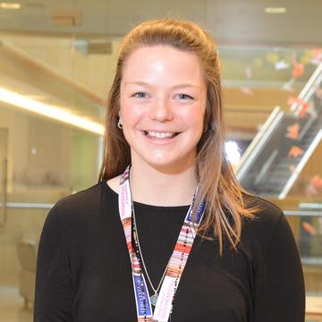
Sarah Silk
Malaria vaccine clinical trials
My research focuses on set up, conduct and immunological analyses of blood-stage malaria vaccine candidate clinical trials. These include first-in-human phase I trials through to vaccine efficacy assessment in the UK and in a number of field sites across Africa. I work very closely with our partners to capacity build, conducting laboratory set up, training and technology transfer of immunological assays to these settings.
Controlled human malaria infection (CHMI) studies are a critical tool for down selection of vaccines. We routinely utilise the blood-stage CHMI model for Plasmodium falciparum here in Oxford, and I recently established this in a malaria endemic setting, Tanzania, for the first time.
Additionally, we have an active Plasmodium vivax programme and have both established the blood-stage CHMI model and conducted a number of trials with our valuable bank of P. vivax inoculum to date.
Through characterisation of the humoral and cellular immune responses in these studies we aim to understand the key factors driving a functional and long-lived response in order to guide vaccine development and optimisation.
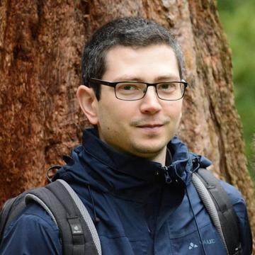
Dr Tim Esser
Cryo-EM of soft-landed β-galactosidase
We have combined soft landing and cryo-EM to obtain a 2.6 Å gas-phase structure of β-galactosidase. Visualizing structural changes on the side-chain level represents a direct, experimental validation of native mass spectrometry. Our workflow forms the foundation for extending the high level of control, selectivity, and robustness of gas-phase methods into more controlled cryo-EM sample preparation.

Dr Suman Dutta
Neurodegenerative disease biomarker assay development
My project focuses on investigating the potential of neuron-originating extracellular vesicles (nEVs) found in human biofluids as diagnostic carriers for neurodegenerative diseases. The absence of exclusive surface markers for central nervous system (CNS) neurons has posed a significant challenge in isolating pure nEVs originating from the brain.
In response to this challenge, my research aims to identify distinct markers on brain-derived nEVs. These markers will enable the precise capture of CNS nEVs from blood samples, potentially transforming minimally invasive liquid biopsies. This advancement promises to enhance the accuracy and accessibility of early disease detection and patient stratification.
Furthermore, the research findings have the potential to deepen our understanding of intercellular communication within the CNS, opening up new avenues for innovative neurological interventions. By employing a combination of experimental and bioinformatic approaches, we seek to achieve these crucial research objectives.

Dr Emma Silvester
DNA nanostructures as tags for cryo-electron tomography.
In my current research project I am developing DNA nanostructures as tags for cryoET. In the Baker lab, we study complex biological systems in 3D by cryo-electron tomography. The challenge lies in identifying specific molecules in crowded environments. The project introduces DNA nanostructures as highly visible and specific protein labels for cryoET. Initially demonstrated on vesicles, viruses and cell surfaces, my research now focuses on intracellular applications. To this end, I am currently designing novel nanostructures to change shape upon successful protein binding to distinguish between bound and unbound tags.
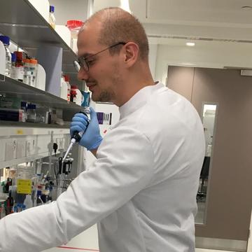
Dr Martin Rieu
Studying the rotary biological motors using gold nanorods and polarization microscopy.
Many species of bacteria propel themselves through the rotation of a flagella of several microns. Rotation is created by a complex nanomachine, the bacterial flagellar motor, which uses the spontaneous flow of some ions (protons, sodium, magnesium depending on the species) through the inner membrane of the bacteria to generate mechanical movement. They do so by converting the chemical energy of ions to mechanical energy with an efficiency close to the best that is allowed by thermodynamics. We use new physical methods to push the boundaries of our understanding of that machine. We attach gold nanorods of a few tens of nanometres to this motor. They scatter light like an electromagnetic dipole, with a very specific pattern and a polarization which depends on their orientation. We then build microscopes to measure the polarization of the light by those antennas and observe the rotation of the biological motor with unpresented time and spatial precisions. This gives us access to a whole new library of biophysical data which shed light on how remarkable physical properties (energetic efficiency, stability, robustness, adaptability) can emerge from complex assemblies of proteins.
Dr Vojta Pražák
Studying bacterial programmed cell death by electron cryo-tomography
Bacteria have multiple programmed cell death pathways, analogous to those found in eukaryotic cells. These are triggered in response to, for example, bacteriophage infection or the presence of antibiotics. I’m developing methods to study bacterial cell death using electron cryo tomography and correlative light and electron microscopy. These methods will allow us to link phenotypic changes to specific structural and molecular mechanisms with the ultimate aim to develop new avenues of treating bacterial infections.

Benjamin Breant
The dissociated state induced by psychedelics.
My research focus on describing the dissociated state induced by the injection of psychedelics in mice. We discovered that the psychedelic we are using increase sleep-like brain activity during wakefulness, so we use novel approaches to characterise levels of arousal.

Dr Abraham Oluwole
Understanding bacterial cell wall biosynthetic enzymes and how they can be intercepted by antibiotics
The world is yet to fully recover from the impact of the Covid-19 pandemic. Yet, antibiotic resistance presents another serious threat to humanity because of its grave clinical and economic impact. Why? Pathogenic bacteria are developing resistance to conventional antibiotics at a pace much faster than the timescale in which new antibiotics are being discovered and approved for clinical use. We want to change that. To achieve this, we are developing new approaches to track core biosynthetic pathways in pathogenic bacteria using mass spectrometry and related techniques. This will allow us to identify new protein targets for existing antibiotics and provide alternative strategies to test and elucidate modes of action of new antibiotic candidates. We focus on membrane proteins that mediate biosynthesis and translocation of bacterial cell wall peptidoglycan, a net-like polymer that provide bacteria with their shape and tolerance to environmental assault. Compromising the integrity of the bacterial cell wall will render them vulnerable to antibiotics and help overcome the ‘silent pandemic’ of antimicrobial resistance.
Dr Stelios Chatzimichail
Development of microfluidic platforms and microscopy techniques for the early detection and AMR testing of pathogens in patient samples.
In this presentation I showcased how we developed a microfluidic platform and molecular assays that enabled us to tell what species is presented in clinical samples and subsequently proceed to phenotype them in order to understand which antibiotics they would most respond to.


