
KINsD post-doctoral researchers present at Interdepartmental Poster Session
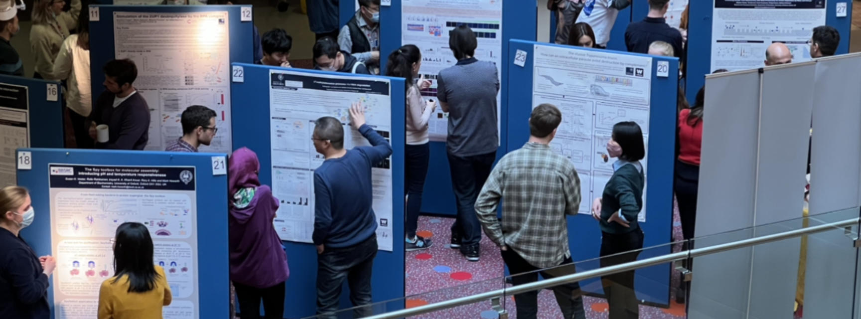
The Department of Biochemisty's Postdoc Society hosted an Interdepartmental Poster Session for neighbouring departments on the South Parks Road science campus, Oxford on Tuesday, 26th April 2022. The event was organised in collaboration with the post-doc societies from the Dunn School of Pathology, the Department of Physiology, Anatomy and Genetics and the Centre for Medicines Discovery.
It was fantastic to see over forty poster presentations from across departments within both the Medical Sciences and the Mathematical, Physical and Life Sciences Divison at Oxford University.
Twenty two Kavli INsD post doctoral researchers presented their recent work at the event held in the beautiful atrium at the Dorothy Crowfoot Hodgkins Building. The building is home to the Department of Biochemistry and the Kavli Institute for Nanoscience Discovery.
Many researchers met for the first time at a drinks reception in the late afternoon to socialise and discuss collaborative opportunities.
Below are the abstracts of the KINsD researchers only. The full document of Poster Abstracts for all departmental participants can be provided on request from info@kavlinano.ox.ac.uk
*Sponsorship received from MERCK and PCR Biosystems
John Young
Combining Peptidiscs and Mass Photometry to characterize membrane proteins in aqueous solution
Carol Robinson, Chemistry / Kavli INsD
Mass Photometry (MP) is an emerging technique for measuring the molecular weight of proteins in solution and for quantifying protein-protein interactions. Key advantages of MP as a tool for protein analysis include its accuracy; it’s speed and ease of use; and its minimal consumption of valuable biological sample. When interfaced with membrane mimetics such as nanodiscs and SMALPs, MP has shown great promise as a method to characterize even the most challenging membrane proteins and complexes.
We here combine MP with peptidiscs – a recently developed membrane mimetic – to measure the molecular weight of membrane proteins in detergent-free conditions. Using two different bacterial ABC transporters, we show that our newly developed peptidisc-MP workflow allows us to visualize interactions between peptidisc-reconstituted membrane protein receptors and their soluble protein binding partners. The effect of nucleotides and other small molecules on these interactions can also be assessed. Using a peptidisc-encapsulated outer membrane receptor, we can visualize and quantify interactions between a membrane protein receptor and a bacteriocidal antibody. In addition to facilitating rapid characterization of receptor-ligand interactions, we believe our peptidisc-MP workflow may also be a promising future tool for screening sample quality prior to structural analysis by X-ray crystallography or by electron microscopy.
Keywords: Mass photometry, Peptidiscs, Membrane protein complexes, ABC transporters

Bethan Cole
Preclinical development of TRPC3 inhibitors to treat spinocerebellar ataxia
Esther Becker, NDCN / Kavli INsD
Spinocerebellar Ataxias (SCAs) are autosomal dominant neurodegenerative disorders affecting the cerebellum, the region of the brain responsible for movement and coordination. Progressive dysfunction of the cerebellum leads to severely impaired quality of life and for many patients is fatal. In addition to problems with balance, speech and movement, patients often display cognitive disabilities, parkinsonism and seizures. There is an unmet need for therapeutics to slow progression of the disorders.
Over 40 pathogenic variants are associated with SCAs, but common mechanisms are thought to underlie different genetic forms. One such mechanism is dysregulated Ca2+ homeostasis in cerebellar Purkinje neurons. TRPC3 regulates Ca2+-signalling and excitability downstream of mGluR1 in Purkinje neurons. Gain-of-function pathogenic variants of TRPC3 have been shown to cause SCA phenotypes both in a patient and a mouse model. Dysregulated TRPC3 signalling is thought to be a pathophysiological mechanism in a number of other SCA subtypes, making TRPC3 a promising therapeutic target.
In collaboration with medicinal chemists, we aim to generate and optimise drug leads to target TRPC3. This is being carried out using a combination of in vitro assays and in vivo preclinical testing methods and we hope to eventually translate these drugs into clinical testing in SCA patients.
Keywords: Spinocerebellar ataxia, Ion channels, TRPC3, Drug development

Jussi-Pekka Tolonen
Pathogenic variants in ITPR1 cluster in functional domains
Esther Becker, NDCN / Kavli INsD
The ITPR1 gene encodes the inositol 1,4,5-trisphosphate (IP3) receptor type 1 (IP3R1), a critical player in intracellular calcium signalling. Pathogenic variants in ITPR1 cause neurodegenerative Spinocerebellar Ataxia Type 15 (SCA15) and congenital SCA29, Gillespie Syndrome, and Severe Pontine/Cerebellar Hypoplasia. The pathophysiological basis of these different phenotypes is poorly understood, limiting therapeutic development. Here we report a time-course analysis of ITPR1 expression and alternative splicing in the human cerebellum, and detailed genotype/phenotype analyses of a large cohort of individuals with pathogenic variants in ITPR1. The individuals were identified using next-generation sequencing as part of the Deciphering Developmental Disorders study, the 100,000 Genomes project, and through clinical collaborations. The gene variants were interpreted using ACMG guidelines. Representing the largest cohort published so far, the data consist of 30 patients with 21 unique variants (eleven novel) that were predicted to be disease-causing based on the concordance of phenotype, the type of mutation present, and correlation with functional analyses reported in the literature. Importantly, the variants cluster in functional domains in the protein, most evidently in the N-terminal IP3 binding domain, and the C-terminal transmembrane channel domain. Our findings have implications for gene-specific variant reporting and can guide ITPR1 variant interpretation in the clinic.
Keywords: Neurodegeneration, Ataxia, Cerebellum, Next-Generation Sequencing, Case Series
Cheng Jiang
Biotechnology Powered Biomarker Discovery for Parkinson's Disease using Extracellular Vesicles
George Tofaris, NDCN / Kavli INsD
Parkinson’s disease (PD) is characterized neuropathologically by α-synuclein aggregation. Currently, there is no blood test using existing biomarkers to predict the underlying pathology or early diagnostics. We investigated the potential clinical utility of serum neuronal extracellular vesicles (EVs) as biomarkers. Due to the low abundance of EVs from the neuronal subpopulation and the complexity of the sample matrix, we developed and set up (1) antifouling immunobeads to specifically capture neuron-derived EVs with the resistance of nonspecific adsorptions, (2) multiplexed assay platform to quantitatively detect internal cargo in a high-throughput format based on bulky measurement, (3) digital droplet microfluidic assay to evaluate the membrane antigens on single-entity EV level. Such platforms have dramatically advanced the progress of PD biomarker study toward translational and personalized medicine.
Benedict Tanudjojo
Phenotypic manifestation of ⍺-synuclein strains derived from PD and MSA in iPSC-derived DA neurons
George Tofaris, NDCN / Kavli INsD
The progressive accumulation and subsequent aggregation of a small neuronal protein, alpha-synuclein, contributes majorly to the symptoms that characterise Parkinson’s disease (PD) and related neurodegenerative disorders. To date, only partially effective symptomatic therapies exist. Therefore, there is an urgent unmet need to inform curative strategies by attempting to answer the question of how does aggregated alpha-synuclein kill neurons? To investigate the mechanisms underlying neuronal death, we generated a model using human neurons derived from patient stem cells, which recapitulated key disease features. Our study demonstrated that the extent of aggregation was dependent on the abundance of alpha-synuclein within neurons—and importantly, that the toxicity of alpha-synuclein was determined by subtle differences in the three-dimensional structure of aggregates. We screened for proteins that interacted with structurally different alpha-synuclein aggregates and discovered 56 candidates, one of which we further explored. In summary, our findings provide an insight into the complex nature of aggregation and how it ultimately leads to the demise of neurons. We define (i) alpha-synuclein levels, (ii) aggregate morphology and (iii) divergent interacting partners as the most important players in our model.
Keywords: Seeded aggregation, iPSC, Synuclein strain

Zihao Wang
cvHsp binds FLNC to compete its dimer interface, and the interaction is regulated by FLNC phosphorylation
Justin Benesch, Chemistry / Kavli INsD
Small heat shock proteins (sHsps) are a family of ubiquitous proteins that defend the first line for proteostasis. They have specific regulatory roles to fulfill their chaperone functions or maintain the physiological structures of tissues through interactions with their native or dysfunctional partners. Among the sHsps, cardiovascular heat shock protein (cvHsp, HspB7) performs its primary functions in cardiac tissues and skeletal muscles. Unlike the well-studied human sHsps forming large oligomers based on dimeric building blocks, we found cvHsp is dominantly monomeric at dynamic conditions. The crystal structure explains that oligomerization and dimerization are much less favored for cvHsp. We proved that dimerized domain 24 (d24) of the actin-linking protein filamin C (FLNC) is the binding substrate of cvHsp. The monomeric cvHsp binds to the dimerized FLNCd24 and competes to its dimerization. Phosphorylation at a threonine residue or a tyrosine residue under different stress conditions has opposite effects in mediating the FLNC dimerization and its interaction with cvHsp. Overall, our study provides new and deep insights into sHsps-regulation in cytoskeleton using a variety of biophysical techniques.
Keywords: Cytoskeleton, Native MS, Crystallography, PTMs

Alex Cook
Invariant surface glycoprotein 65 of Trypanosoma brucei is a complement C3 receptor important for virulence
Matthew Higgins, Biochemistry / Kavli INsD
African Trypanosomes replicate in the blood of mammalian hosts yet are completely exposed to the adaptive and innate immune systems. Despite this, Trypanosomes can sustain long-term infections - how can this parasite survive constant host immune-surveillance? Trypanosomes evade the adaptive immune response through antigenic variation of a surface coat consisting of a dense layer of variable surface glycoprotein. However, very little is known about how they negate the innate system, including the blood circulating complement system. We have discovered that an invariant surface glycoprotein, ISG65, is a receptor for Complement Component 3 (C3). We show how ISG65 binds to the thioester domain of C3b. We also show that knockout of the ISG65 locus greatly decreases the pathogenicity of trypanosomes in a mouse model. Deposition of C3b on pathogen surfaces is a central point in activation of the complement system and C3b has been observed on trypanosome surfaces. Our findings therefore suggest that trypanosomes have a C3 receptor distributed across their surfaces that greatly decreases their susceptibility to complement-mediated killing.
Keywords: Infectious disease, Complement system, Trypanosome, Structural biology
Barney Williams
Monoclonal antibodies against Plasmodium falciparum RH5-Interacting Protein (RIPR) inform blood-stage malaria vaccine design
Simon Draper, Biochemistry / Kavli INsD
The development of blood-stage malaria vaccines has been hampered by a lack of conserved antigens capable of inducing cross-strain neutralising antibodies. The most promising blood-stage candidate to date is RH5, currently being evaluated in clinical trials. RH5 forms the RCR complex with Plasmodium falciparum RH5-interacting protein (RIPR) and cysteine-rich protective antigen (CyRPA). RIPR is a ~120kDa protein comprising 10 Epidermal Growth Factor (EGF)-like domains. We generated anti-RIPR polyclonal antibody (pAb) by immunising rabbits with full-length RIPR protein. This pAb effectively inhibited cultured parasites in a standardised Growth Inhibition Activity (GIA) assay. Recombinant RIPR protein fragments were used to reverse GIA of the anti-RIPR pAb. Using GIA reversal we determined that the neutralising epitopes are located on a 184 amino acid region of RIPR corresponding to RIPR EGF(5-8). The importance of EGF(5-8) was confirmed by the generation of GIA positive monoclonal antibodies (mAbs) only against this region. All neutralising mAbs were able to bind, but not disrupt the RCR complex. In contrast, non-inhibitory mAbs could prevent in vitro formation of the RCR complex indicating that disrupting complex formation is not the mechanism of parasite invasion inhibition. These data were used to design next-generation vaccines containing the RIPR antigen.
Keywords: Malaria, Vaccine, Antibody
Bevin Gangadharan
Novel biomarkers for fatty liver disease
Nicole Zitzmann, Biochemistry / Kavli INsD
Approximately 1 in 3 people in the UK have some degree of non-alcoholic fatty liver disease (NAFLD) and most affected individuals are unaware they have the disease. Currently, the reference standard for assessing NAFLD is the liver biopsy, an invasive, painful procedure which can be unreliable and costly. Consequently, there is a need for improved, minimally-invasive methods of determining stages of NAFLD in patients.
Mass spectrometry (MS) and two dimensional gel electrophoresis (2DE) were used to separate proteins in serum from patients with NAFLD. Several potential biomarkers were identified and both Western blotting and targeted MS were used to check these biomarkers. An antibody-free assay was developed for one of our most promising NAFLD biomarkers (apolipoprotein F) using parallel reaction monitoring. Our biomarkers look promising when compared to other NAFLD biomarkers currently used in the clinic.
The novel biomarkers would allow clinicians to identify NAFLD patients so they could be advised about lifestyle changes, such as diet and exercise, which could help to reverse the disease and thus save money for healthcare providers. The technology could be used to diagnose, prognose or monitor the progression of NAFLD in an individual thereby reducing the need for a liver biopsy.
Keywords: Proteomics, Mass spectrometry, LC-MS, Biomarkers, Fatty liver disease

Helen Rowland
Characterising inflammatory response in iPSC-derived astrocytes
Noel Buckley, Psychiatry / Kavli INsD
Neuroinflammation plays an active role in progression and development of Alzheimer’s disease (AD). Under neuroinflammatory conditions astrocyte function is affected and dependent largely on the sub-populations involved, and the context of the inflammation. In this study, we seek to determine how different inflammatory stimuli affect iPSC-derived astrocytes, and if deep learning methods can be used to distinguish stimuli.
iPSCs derived from patients with AD were differentiated for 100+ days to cortical astrocytes. Astrocytes were then treated with a range of different neuroinflammatory stimuli to induce reactivity. Using immunocytochemistry astrocytes were imaged for general astrocytic and neuroinflammatory markers on the Opera Phenix high-content imaging system. Images were then analysed via Harmony analysis software to measure changes in protein expression and morphology. This was compared to deep learning approaches.
Preliminary data demonstrated some neuroinflammatory treatments caused distinguishable changes in protein expression and cell morphology and that these effects varied between patient lines. This analysis was surpassed by the deep learning approaches which could more accurately distinguish treatment types.
Deep learning approaches offer new insight into classification of astrocytes and neuroinflammation. Our findings demonstrate a potential novel method for screening therapeutics in Alzheimer’s disease.
Keywords: iPSC, Astrocyte, Alzheimer's disease, Inflammation

Stephen Thorpe
Combining escape time electrometry with mass photometry for protein separation by mass and charge
Philipp Kukura, Chemistry / Kavli INsD
Diseases such as triple negative breast cancer or glioblastoma have heterogenous tissue responses that must be probed across length scales, from whole tissue to single cell. Single cell proteomics lacks the dynamic range to measure biologically relevant low abundance proteins and cannot measure multiple combinations of post transcriptional modifications. New proteomics technology is required to meet these challenges. Mass photometry is a label free method of measuring the mass of single molecules in solution. Escape time electrometry measures the charge of a single molecule confined within a nanoscale electrostatic trap, with sub electron charge sensitivity. We propose using ETe as a charge-based separation step and MP as the detection modality allowing single molecule label free measurement of both mass and charge in a method analogous to ion mobility mass spectrometry. Using a new ETe trap geometry we demonstrate separation of 30,60 and 120 base pair double stranded DNA. We have theoretically investigated nanofluidic sample injection capable of generating sub 100 ms pulses for synchronous delivery of samples and have integrated this into a glass nanofluidic device compatible with MP. We characterise the devices accuracy and resolution for quantifying both mass and charge using known protein ladders.
Keywords: Nanofluidics, Label free mass photometry, Electrometry separation
Elena Britti
Phenocopying mitochondrial Parkinson’s disease patient phenotypes using CRISPRi in iPSC-derived neurons
Richard Wade-Martins, DPAG / Kavli INsD
Parkinson’s disease (PD) is a neurodegenerative disorder characterized by dopaminergic neurons loss in the substantia nigra pars compacta. Point mutations in α-synuclein (SNCA), PTEN-inducible kinase-1 (PINK1) and Parkin ubiquitin E3 ligase (PRKN) genes, but also SNCA triplications, have been identified to cause PD. Characteristics of PD are α-synuclein aggregation and mitochondrial dysfunction. Dysfunctional mitochondria are marked for mitophagy by PINK1 and Parkin with phospho-serine65ubiquitin (pS65Ub).
Our lab has developed novel PD models using induced-pluripotent stem cell (iPSC)-derived dopaminergic/cortical neurons and CRISPR interference (CRISPRi). CRISPRi allows sequence-specific repression of gene expression by using sgRNAs to downregulate genes such as PINK1 and PRKN. Using CRISPRi, we recapitulated patient cell phenotypes such as pS65Ub loss, as seen in iPSC-derived dopaminergic neurons from patients. We aim to use it in combination with SNCA triplications to analyse gene repressions in another context. Our data indicate that CRISPRi technology can phenocopy patient-derived cells to understand the mechanisms of PD to identify new therapeutic approaches.
Keywords: Parkinson's disease, Mitochondria, CRISPR interference, Phospho-Serine65 Ubiquitin
Sam Tusk
An Introduction to Mass Photometry – Label-Free Mass Measurement in Solution
Philipp Kukura, Chemistry / Kavli INsD
Mass Photometry is a technique which uses interferometric imaging to measure light scattered by single biomolecules as small as 30kDa, without the need for labels. For proteins (and protein complexes) the amount of scattered light is proportional to the molecule’s mass, allowing different species to be distinguished and identified.
In one configuration, proteins are measured as they adsorb onto a glass surface from solution. The resulting measurements reflect the mass distribution of the proteins in solution, in their native state. This can be used to characterize biomolecular interactions, or to characterize sample purity.
In a different configuration, biomolecules are measured as they diffuse across a glass surface (e.g. when embedded in a supported lipid bilayer). Both mass and diffusion coefficient are measured over time.
The Kukura lab is focussed on pushing the technical limits of Mass Photometry, and on expanding its application to different types of assays and different types of biological systems. We are also committed to making Mass Photometry accessible – in the Kavli institute we host a Mass Photometry facility with a number of simple-to-use commercial instruments, and we are always open to collaborations.
Keywords: Label-free, Mass distribution, Mass photometry, Protein characterization, Single-molecule
 presenting on behalf of Sam Tusk.")
Jack Peters (DPhil student) presenting on behalf of Sam Tusk
Jan Becker
Revealing the basics of mass photometry with Fourier optics
Philipp Kukura, Chemistry / Kavli INsD
Mass photometry has recently emerged as a quantitative approach to study biomolecular structure, dynamics and interactions at the single molecule level without the need for labels. Here, we employ Fourier optics to model light scattering from nanoscopic objects and image formation by interference with background light. Coupled with atomic force microscopy of glass substrates commonly used in mass photometry, we obtain a theoretical description of mass photometry in close agreement with experiment in terms of both imaging background and commonly encountered signal magnitudes for polypeptides with appropriate mass scaling. These results enable a quantitative understanding of mass measurement by light scattering, and open up avenues for in silico exploration for improving detection sensitivity and improving measurement precision for next generation mass photometry.
Keywords: Mass photometry, Fourier optics, Simulation, Interference scattering microscopy, In-silico

Anna Olerinyova
Mass Photometry of Membrane Proteins
Philipp Kukura, Chemistry / Kavli INsD
Integral membrane proteins (IMPs) are biologically highly significant but challenging to study because they require maintaining a biological lipid-like environment. Here, we explore the application of mass photometry (MP) to IMPs and membrane-mimetic systems at the single-particle level. We apply MP to amphipathic vehicles, such as detergents and amphipols, as well as to lipid and native nanodiscs, characterizing the particle size, sample purity, and heterogeneity. Using methods established for cryogenic electron microscopy, we eliminate detergent background, enabling high-resolution studies of membrane-protein structure and interactions. We find evidence that, when extracted from native membranes using native styrene-maleic acid nanodiscs, the potassium channel KcsA is present as a dimer of tetramers—in contrast to results obtained using detergent purification. Finally, using lipid nanodiscs, we show that MP can help distinguish between functional and non-functional nanodisc assemblies, as well as determine the critical factors for lipid nanodisc formation.
Keywords: Mass photometry, Membrane proteins, Detergents, Lipid nanodiscs, Native nanodiscs

Manish Singh Kushwah
Understanding Dynamin Polymerization by Single-molecule Counting and Particle-tracking Using Mass Photometry
Philipp Kukura, Chemistry / Kavli INsD
Dynamin, the prototypical member of the dynamin-superfamily of membrane-remodelling proteins, catalysed membrane fission critically relies on its polymerization. Here, we use label-free, mass photometry (MP) to understand the dynamics of dynamin oligomers in both solution and on membranes. We find that in solution, dynamin is a polydisperse protein
and dimer and tetramer are the most abundant species at physiological salt concentration. Relative abundance of various oligomers is determined by the bulk protein concentration, and the GTPase cycle. Using a quantitative assay to study dynamin polymerization with MP, we compared the dissociation constant (Kd) for all oligomers in non-polymerizing and polymerizing conditions and find that both tetramer and dimer coordinate to control dynamin polymerization where tetramer and dimer function as the nucleating and the repeating unit, respectively. Furthermore, we studied mass and diffusion of dynamin oligomers via label-free single-particle tracking using MP on supported bilayers and find that membranes induce dynamin polymerization by relieving pH-domain mediated autoinhibition. We further show that the membrane mediated regulation is uncoupled in centronuclear myopathies (CNM) mutants leading to significantly higher polymerization in solution and higher effective affinity to membranes. Taken together, we describe the mechanism of dynamin polymerization and its regulation in solution and on membranes as well as provide a template to quantitatively study polymerization of membrane binding proteins.
Keywords: Dynamin, Protein-protein interactions, Polymerization, Single-particle tracking, Membrane affinity

Roi Asor
The Spike-ACE2 interaction underlying SARS-CoV-2 infection and inhibition is enhanced by intermolecular crosslinking
Philipp Kukura, Chemistry / Kavli INsD
The interaction between the SARS-CoV-2 spike trimer with the host dimeric angiotensin-converting enzyme 2 (ACE2) receptor is the first step in virus entry. Consequently, efforts aimed at therapeutic intervention have focused on blocking spike-ACE2 binding by designing ACE2-based decoys. The multivalency of ACE2 and spike is fundamental to our understanding of the molecular mechanism underlying ACE2-spike affinity, manifested by the observation of much tighter binding of dimeric ACE2 to spike compared with monomeric ACE2. While structural evidence suggests that intra-spike avidity is unlikely to explain these observations, the alternative, intermolecular avidity mechanism through ACE2-mediated crosslinking is challenging to detect and quantify owing to the inherent heterogeneity of crosslinked oligomeric complexes.
Using mass photometry, we quantified the free energies for the spike-ACE2 interaction building blocks; the spike receptor binding domain and soluble monomeric or dimeric ACE2. Similarly, we quantified the interaction between monomeric ACE2 and trimeric soluble spike.
For dimeric ACE2 and trimeric spike, we found that the mass balance was shifted, predominantly favoring high-mass oligomers with increasing ACE2 concentration. Our results show that in solution, ACE2 dimer and spike trimers can crosslink, which coupled with the enhanced inhibitory effect of dimeric ACE2 compared to monomeric ACE2 on viral infection, suggests that inter-molecular ACE2-spike avidity maybe the underlying mechanism enhancing the spike-ACE2 interaction.
Keywords: Mass-photometry, Label free single particle detection, Protein-protein interactions, COVID

Emanuel Pfitzner
Fluorescence-assisted mass photometry
Philipp Kukura, Chemistry / Kavli INsD
Total internal reflection fluorescence microscopy can detect single fluorescently labeled molecules close to a surface. Despite its high specificity, the fluorescent probes need to be attached the individual proteins. Mass photometry (MP) surpasses this limitation by interferometrically detecting elastic scattering which allows the localization and mass measurement of single proteins. MP, however, struggles with detecting species of low molecular mass (< 40 kDa) and its non-specificity prevents particles of similar mass to be distinguished solely by the scattering readout.
Here, we report on the combination of single molecule fluorescence and scattering microscopy to improve the specificity and sensitivity of mass photometry. Recording landing assays of different fluorescently labeled proteins, we were able to differentiate molecules carrying a fluorescent tag from molecules without the tag in a quantitative manner even though they would be detected at a very similar mass, thus, drastically increasing MPs specificity.
To test the improvement in sensitivity, we used an intrinsically fluorescent protein as a probe: green fluorescent protein (GFP, 27 kDa). The high specificity of GFPs fluorescence emission allowed us to locate it in time and space during a landing event. The knowledge about the proteins position significantly improved the detection limit of MP.
Keywords: Mass Photometry, Single-molecule

Remya Raghavan Nair
Mitophenotyping of primary neuronal culture models of Parkinson’s disease
Richard Wade Martins, DPAG / Kavli INsD
Parkinson’s disease (PD) is a progressive neurodegenerative disease characterised by loss of dopaminergic neurons (DANs) in the substantia nigra region of the mid-brain. Mutations in SNCA and LRRK2 genes are reported to be associated with autosomal dominant forms of familial PD. Mitochondrial dysfunction is involved in the development of pathological changes in DANs in PD. Studying the role of mitochondrial dysfunction in the pathogenesis of PD is important for the development of therapeutic strategies.
To visualize the mitochondrial architecture and mitophagy in cell culture models of PD, we used mitoQC tool. We have generated rat cortical cell culture carrying R1441C mutation in LRRK2 gene and transfected it with mitoQC reporter. The mouse line carrying SNCA over expression (SNCA-OVX) displayed clear mitochondrial dysfunction in mid-brain. To study the mitophagy and mitochondrial morphology, we obtained primary cortical neurons from a double transgenic mouse line carrying both the SNCA-OVX and mitoQC transgenes. Both rat and mouse primary cortical cultures showed a clear reduction in mitophagy by high throughput imaging analysis. Moreover, the mitochondrial morphology was altered in primary cortical cultures from SNCA-OVX line. These data indicate that both models are powerful tools to study the PD mitochondrial phenotype.
Keywords: Parkinson's disease, Neurons, Mitophagy

Gizem Önal
The effects of GBA mutation severity on α-synuclein degradation mechanisms in iPSC-derived dopamine neurons
Richard Wade Martins, DPAG / Kavli INsD
Mutations in the GBA gene are the most common genetic risk factor for Parkinson's Disease (PD). The 'mild' (N370S) and/or 'severe' (L444P, D409H) GBA mutations are known to have differential effects on the development of PD, including age-of-onset, clinical symptoms, and disease severity. In this study, I investigated the effects of GBA mutation severity dependent molecular mechanisms underlying the role of α-synuclein degradation mechanisms. N370S, L444P, and D409H GBA mutant iPSC-derived dopamine neurons were used to analyze the degradation of a-synuclein via ubiquitin-proteasome system, chaperone-mediated autophagy (CMA), and macroautophagy. The immunostaining and immunoblotting assays were used to measure the localization and protein levels of a-synuclein, ER-stress and autophagy-lysosome pathway (ALP)-associated proteins. The release of α-synuclein was analyzed by Quartz Crystal Microbalance with Dissipation (DQCM) monitoring. I found that 'severe' GBA mutations triggered significant accumulation and release of α-synuclein in iPSC-derived dopamine neurons. ER-stress and total ubiquitination levels were significantly higher in severe mutant GBA lines compared to mild ones. Impairments in CMA were detected in 'mild' GBA-mutant line, whilst macroautophagy was disrupted in ‘severe’ GBA mutant lines. Overall, these results may help to explain the molecular basis of the differential clinical PD features based on GBA mutation severity.
Keywords: GBA, Parkinson's disease, α-synuclein, ER stress, Autophagy

Sean Burnap
The influence of stabilising mutations upon SARS-CoV-2 spike glycan processing, dynamics and function
Weston Struwe, Chemistry / Kavli INsD
The severe acute respiratory syndrome (SARS)-coronavirus (CoV)-2 is decorated with trimeric spike glycoproteins that mediate binding to host cells. The structural study of these spike complexes is hindered by proteolytic cleavage and inherent instability, resulting in the transition from a prefusion to the more stable postfusion state. The introduction of several proline substitutions within spike have enabled a dramatic stabilisation of the prefusion state, increasing protein yield, to act as a superior immunogen. Furthermore, the glycosylation state of spike greatly influences antibody recognition and is therefore of paramount importance for vaccine design. However, it remains unclear how stabilisation modulates glycan processing, and the extent to which these alterations influence spike function. Utilising mass photometry we reveal that the extent of proline substitutions used for stabilisation directly hinders the ability of SARS-CoV-2 spike to bind its host receptor angiotensin-converting enzyme 2 (ACE2), which was consistent among Wuhan and Omicron variants. Glycomics and glycoproteomic analyses highlighted no major change in glycan processing upon spike stabilisation, suggesting that differences in ACE2 binding are not related to glycan structure. Further work utilising hydrogen deuterium exchange-mass spectrometry will aim to uncover the structural basis as to how spike stabilisation alters structural dynamics and ACE2 binding.
Keywords: SARS-CoV-2, Spike Glycoprotein, Protein Stabilisation, Glycans, Mass Photometry

Martino Bardelli
Viral vectored delivery of monoclonal antibody genes against blood-stage malaria
Simon Draper, Biochemistry / Kavli INsD
A significant challenge in the development of a malaria vaccine is the lack of antigen formulations that induce the high concentrations of functional antibodies required to achieve protection. Using potent monoclonal antibodies (mAbs) as prophylactics, combined with anti-malarial drugs, could bypass the need for a vaccine to aid in current malaria elimination programmes, but the high cost of this technology makes it incompatible within a developing world context. Vectored immunoprophylaxis (VIP) could provide a cost-effective strategy to implement this vision. This approach uses viral vectors such as adeno-associated virus (AAV) to deliver mAbs that are expressed in situ into the plasma from virally-transduced cells following intramuscular immunisation.
Keywords: Monoclonal antibodies, Malaria, VIP, AAV
